
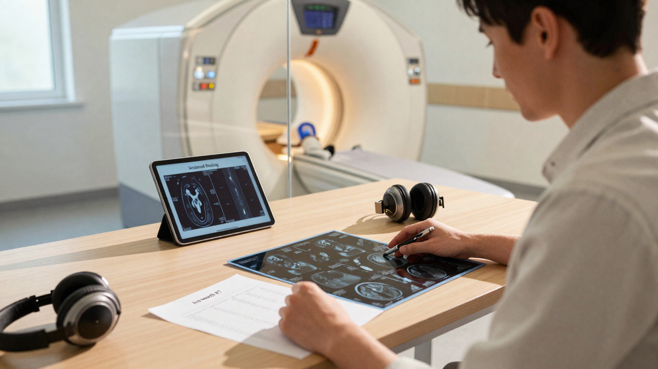
For a considerable price, many private clinics across the UK provide full-body MRI scans intended to identify disease and abnormalities as early as possible. Kim Kardashian and Kate Hudson are among their supporters. (Other celebrity endorsements are available.)
But how do these scans function, and are they really "life saving", as Kardashian claims?
How full-body MRI scans work
MRI generates highly detailed, three-dimensional anatomical images of nearly every internal part of the body, including organs, bones, muscles and blood vessels. The scanners use a powerful magnet and radio waves to produce these body images.
In contrast with X-rays and CT scans, MRI does not use ionising radiation, which can raise cancer risk by a very small amount.
In the UK, a full-body scan costs between £1,000 and £3,000. Although these scans offer a useful general picture of a person's anatomy, they are not tailored to provide the best possible view of every organ or bodily system.
If anything unusual is identified, it would require additional investigation using specialist MRI scans, for example of the heart or prostate. This is because the scanner must be set to different parameters to achieve optimal imaging for particular organs and systems.
It is also important to recognise that MRI scans cannot identify most preventable illnesses. Heart disease, high blood pressure, high cholesterol and diabetes account for the majority of deaths.
MRI scans cannot diagnose these conditions. While they might reveal certain structural changes, other signs are likely to have appeared beforehand.
Incidental findings on MRI scans
Full-body MRI scans can reveal abnormalities known as "incidentalomas", so called because they are chance findings on scans carried out for other symptoms.
Research examining more than 16,000 volunteer brain MRI scans found that fewer than 4 percent included abnormalities needing further investigation.
Brain scans can identify pituitary abnormalities, including non-cancerous growths. Reduced blood flow to the brain without symptoms has been found in 7 percent of people, while aneurysms have been detected in 1.8 percent and benign tumors in 1.6 percent of the population.
Cavernomas, which are small groups of abnormal blood vessels, seldom produce symptoms and appear on MRI scans in around one in 600 people. Symptomatic cavernomas are found in roughly one in 400,000 people.
The spine is another part of the body where incidental findings are common. MRI is increasingly used for back pain, which can uncover incidentalomas such as harmless bone growths and hemangiomas: small blood-vessel growths located on or within the spine.
Hemangiomas occur in more than 40 percent of people having MRI scans, and 99 percent cause no symptoms or need for treatment. The 1 percent who experience symptoms, including pain, numbness or weakness, need surgery to stop progression towards paralysis.
Tarlov cysts are protrusions around nerves that leave the spinal cord in the sacral area, close to the base of the spine. They are present in around 4 percent of the population, with most people having no symptoms.
However, people who develop symptoms such as pain and numbness may require surgery to avoid bone deterioration or damage to the nerves supplying the legs, pelvic structures and genitals.
What about serious findings?
A meta-analysis, which combines information from multiple studies, indicates that serious issues detected unexpectedly on MRI scans occur in 1.4 percent of brain scans, 1.3 percent of chest scans and 1.9 percent of abdominal scans.
There is also the possibility of a false positive: an apparent sign of disease that is not actually disease. The breasts, with up to 97 false positives per 1,000 scans, and the prostate, with 29 per 100 scans, are especially prone to false-positive findings.
These scans can provide useful insight into what is happening beneath the skin. Yet many people find the reports and their terminology difficult to understand.
There is substantial evidence that the typical person has difficulty interpreting common medical language, often giving a phrase the opposite meaning to that intended by the doctor, even for one diagnosis. Reports from these scans cover the entire body and therefore include far more medical information than a report on a single diagnosis, including when everything is normal.
When people pay for scans but lack the means to fund follow-up medical tests, already overstretched healthcare systems must deal with the consequences and confirm that false positives and benign growths are indeed harmless.
For people able to afford them, these scans provide a chance to look inside the body, but the probability of discovering something that requires urgent medical care is low. If I had the money for one, I would spend it on a holiday instead, which would be much better for my health.
Adam Taylor, Professor and Director of the Clinical Anatomy Learning Centre, Lancaster University
This article is republished from The Conversation under a Creative Commons license. Read the original article.





Comments
No comments yet. Be the first to comment!
Leave a Comment