
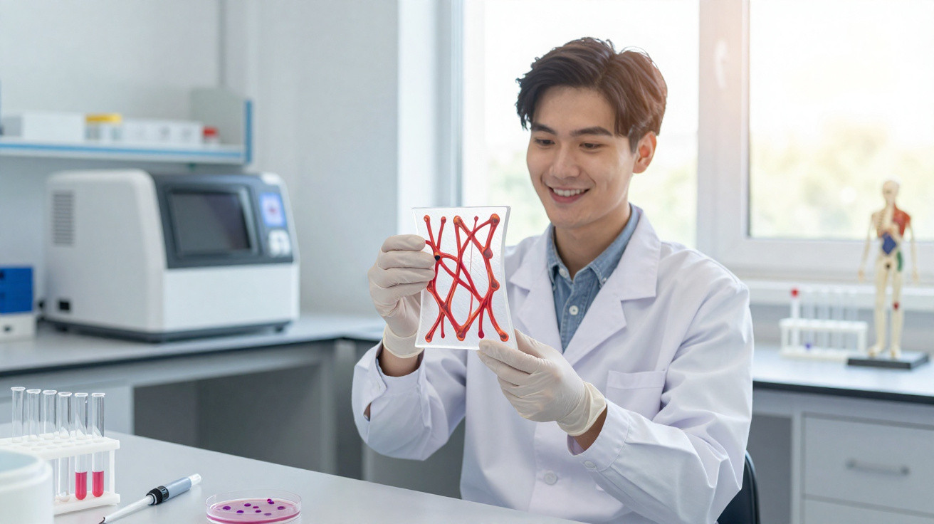
A significant step forward in tissue reconstruction has shown that bone, cartilage, muscle and fat-like tissue can be regenerated using bone marrow stem cells.
The work illustrates the progress made in laboratory-grown repair materials, while placing greater emphasis on whether these constructs can remain viable within the human body.
From cells to shaped tissue
Within 3D-printed scaffold structures, the engineered tissues developed precise forms corresponding to damaged biological structures.
A team at the National Polytechnic Institute (IPN), led by Jorge Vela Ojeda, demonstrated that bone marrow stem cells can be directed to develop into several types of tissue.
Rather than forming one uniform mass, the cells produced separate structures resembling bone, cartilage, muscle and fat.
However, this difference introduces a further challenge: tissue shaped under controlled laboratory conditions is not necessarily able to survive or integrate after implantation.
Multiple outcomes of stem cells
Mesenchymal stem cells are central to the project. This bone marrow cell population can develop into bone, cartilage and fat when exposed to the appropriate signals.
They differ from blood-forming stem cells because they are non-haematopoietic: they belong to the tissue system that supports bone marrow rather than the system responsible for producing blood.
Researchers are interested not only in their developmental potential, but also in the repair signals they release near injured tissue.
This combined ability to build tissue and send signals helps explain their continued role in repair research.
Harvesting from original sources
The study uses material obtained from bone marrow, the soft tissue found within bones that contains several stem cell populations.
According to Vela, the most straightforward collection method is needle aspiration from the iliac crest, the upper edge of the pelvis.
Only a limited amount of material is naturally available at that site. The IPN team says, however, that it can be expanded in the laboratory before being used.
By multiplying these scarce cells, a very small sample can become large enough for testing.
Scaffold structure support
After expansion, the cells were placed onto scaffolds: 3D-printed supports that provide developing tissue with both a shape and a surface to attach to.
Instead of cultivating an unstructured mass, researchers could tailor a construct to a persistent fracture or another damaged area.
Their aim was to produce bone, connective tissue and muscle suited to a non-healing fracture or a particular organ.
In this context, structure is not merely aesthetic, as geometry may determine whether repaired tissue integrates with the body or fails when placed under stress.
Healing signals at work
Repair relies on more than cells establishing themselves and becoming permanent parts of the tissue. Medical interest is also focused on the proteins and tiny vesicles released by these cells, particularly those that reduce inflammation and support the formation of new blood vessels.
This is important because an injured area may first require a more favourable healing environment before it can rebuild itself.
Nevertheless, a construct that performs well in a dish may act differently once blood flow, immune signals and physical forces are involved.
The challenge of consistency
Before an implant can be used in a patient, it must pass a far less glamorous requirement: disciplined manufacturing.
Cells maintained in culture for too long may mutate, shift towards an unsuitable identity or grow in unintended ways.
Regulators require sterility, purity, consistent behaviour and proof that the product will not cause further harm following implantation.
Although these requirements slow progress, they distinguish credible regenerative medicine from hopeful marketing.
Guardrails for new therapies
International guidance makes clear that promising laboratory findings involving complex cell products should not be moved directly into routine care.
Guidelines from the International Society for Stem Cell Research (ISSCR) require safety and effectiveness to be demonstrated in clinical trials before standard use.
Long-term monitoring may also be needed, as transplanted cellular products can remain in the body and cause problems later.
Consequently, the team’s next stage - using the approach with patients with IMSS support - will be considerably more demanding and difficult.
Knowledge from the clinic
Experience also influences how quickly a project of this kind can progress. During 23 years leading haematology at a specialist hospital in Mexico City, Vela has encountered numerous laboratory results that did not succeed.
“It will help this field develop much faster,” said Ojeda.
That potential still relies on stronger experiments and clear evidence, rather than automation alone.
Competing in regenerative medicine
This research has brought Mexico into the field, although Vela noted that the United States, Spain, England and Germany have made the greatest advances.
Regenerative medicine moves forward when biology, materials, surgery and regulation all make progress.
The IPN result is significant because it connects a public university, a medical school and a national health system around tissue injuries that do not heal.
Whether this collaboration develops into a therapy will rest on reproducible results, rather than the apparent scale of the early milestone.
The principal challenge is no longer converting bone marrow cells into replacement tissue, but bringing that tissue safely into clinical practice.
If researchers can close that gap through clean manufacturing, trials and proven follow-up, regenerative medicine in Mexico has a bright future.






Comments
No comments yet. Be the first to comment!
Leave a Comment